
You are currently viewing only those items made visible to the public. Click here to sign in and view the full catalogue.

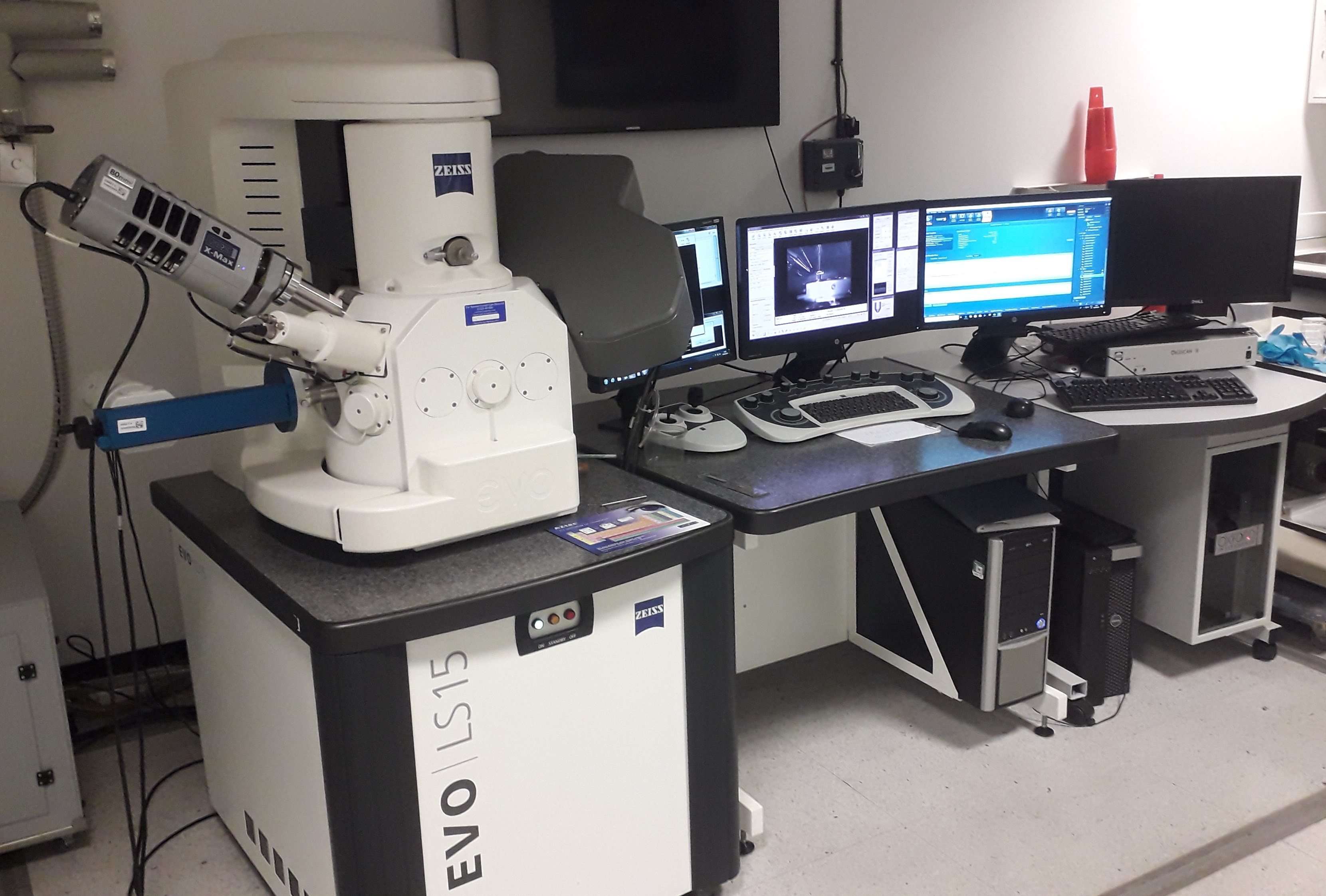
Scanning Electron Microscope
| MANUFACTURER | Zeiss |
|---|---|
| MODEL | EVO 15LS |
| ACRONYM | SEM-EDX |
| AVAILABILITY | Weekdays |
|---|---|
| TRAINING | Training is required to use this item and we can arrange this if needed. |
| CONTACT 1 | John Spratt |
|---|---|
| CONTACT 2 | Tobias Salge |
| Enquire about this item |
Description
The Zeiss EVO 15LS analytical scanning electron microscope (SEM) is a versatile instrument suited to diverse imaging and analytical functions.
The Zeiss EVO 15LS is used for a very wide range of applications, particularly the initial examination of samples. Its user-friendly interface makes it easy to use, even for novices. As little as half a day's training is needed for confidence in most of the common applications. Applications
Imaging the polished and carbon-coated surface of rocks and minerals This is the most frequent application, carried out before detailed composition analysis is performed. Backscattered electron images (BEI) are the most common pictures taken. They show variation in the sample's composition as different grey tones. Composition analysis can then be performed using the Cameca SX100 electron microprobe. Imaging fine surface detail Can be used to image conductive or metal-coated specimens, including biological ones. Looking at the complex surface of samples that are large, rough and unprepared Ideal for examining important museum specimens where their preservation is vital as it doesn’t damage their surface. Creating stereo pair images Can be used for colour anaglyphs or even 3D models whose size and shape can be measured. Identifying minerals The energy dispersive X-ray (EDX) detector is routinely calibrated for analysis of major elements in silicates (the most common rock-forming minerals) and can very rapidly give information necessary for mineral identification. Analysing composition The microscope can be controlled automatically by the EDX detector computer and can be programmed to make maps of large areas of the sample. These maps show differences in composition across centimetre-scale areas and allow measurement of areas. Analyses can be taken at preselected locations, and the programme can hunt for particles of interest. If elements are present at very low concentrations or the EDX cannot separate their signals, the wavelength dispersive X-ray (WDX) detector can be used to create spectrum scans. The Cameca SX100 electron microprobe also has WDX detectors. Cathodoluminescence imaging The cathodoluminescence detector can be used to create images from light emitted by a sample. The images can be used to gain information about the history of mineral deposition and crystal growth
Specification
Stub-mounted specimens, or polished blocks and sections are preferred for this instrument.
Item ID #.
Last Updated: 11th January, 2023
There are no publically available categories listed at present. You may have to sign in to browse this catalogue.
