You are currently viewing only those items made visible to the public. Click here to sign in and view the full catalogue.

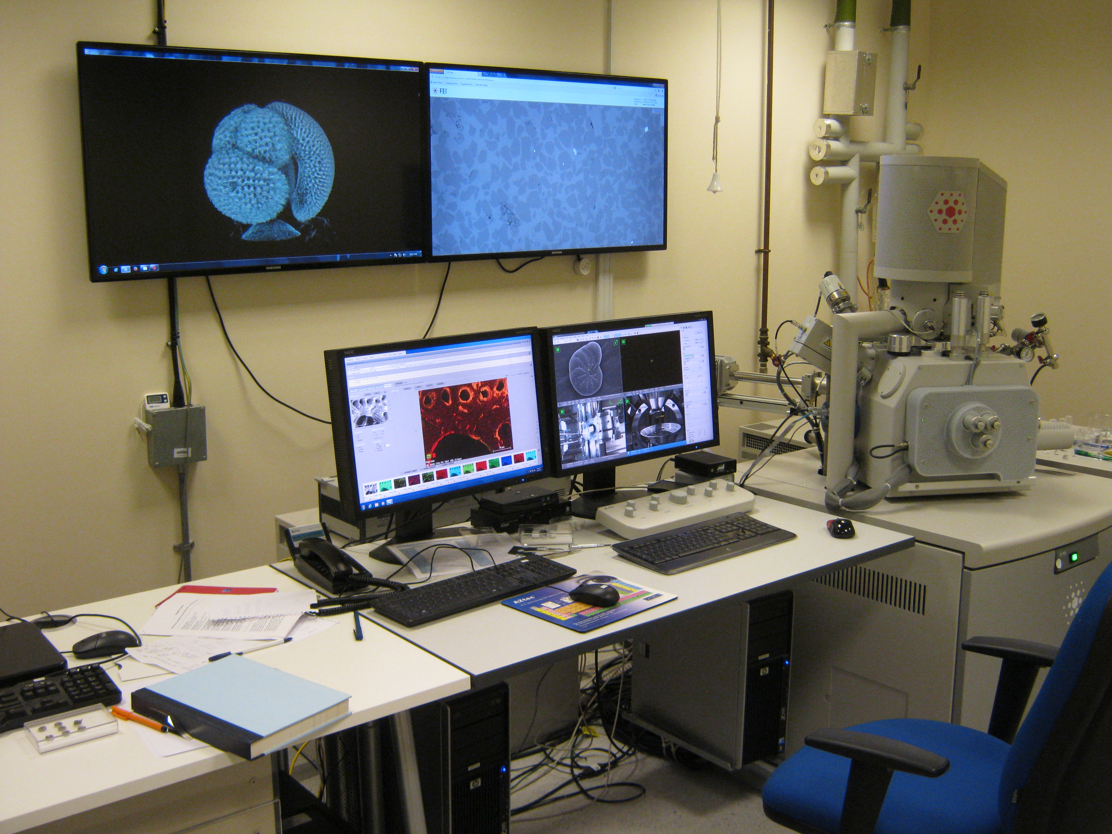
Scanning Electron Microscope
| MANUFACTURER | FEI |
|---|---|
| MODEL | Quanta 650 FEG |
| ACRONYM | SEM-EDX |
| AVAILABILITY | Weekdays |
|---|---|
| TRAINING | Training is required to use this item and we can arrange this if needed. |
| CONTACT 1 | Innes Clatworthy |
|---|---|
| CONTACT 2 | Alex Ball |
| Enquire about this item |
Description
The FEI Quanta 650 FEG scanning electron microscope (SEM) is used for high-resolution imaging and semi-quantitative X-ray microanalysis of both conductive and non-conductive specimens at nanometer resolution.
The FEI Quanta 650 FEG has two modes:
Variable pressure (VP) mode (pressure range 100-200Pa) Used for high-resolution imaging of non-conductive samples in both secondary electron and backscatter modes. High-vacuum (HV) mode (pressure range 10-2-10-4 Pa) Used for high-resolution imaging of conductive samples in both secondary electron (SE) and backscattered (BSE) modes.
It is equipped with the following accessories and features:
a low-vacuum cone, mounted on the final lens, for low-kV imaging under VP conditions to improve imaging of surfaces, with reduced beam penetration a beam sleeve, mounted on the final lens, to improve the performance of Energy Dispersive X-ray micro-analysis (EDX) in VP conditions by reducing beam skirt beam-deceleration (BD) mode for ultra-high resolution backscatter imaging of flat samples under HV conditions drift-correction mode for imaging highly charging samples MAPS software for tiling and creating large montage images at high resolution, which allows for automated image collection of large areas or multiple areas custom-built vacuum pumping system allowing for slow and fully controllable pump down time for specimens sensitive to rapid decompression rates additional micro tilt/rotate stage (Deben) for high-precision work on small, demanding samples, e.g. pinned insects Bruker Flat Quad 5060F energy dispersive X-ray detector (EDS) for ultra-fast and shadow-free semi-quantitative elemental analysis and hyperspectral mapping, even on samples that are non-conductive and highly topographic
Specification
Sample sizes can range from standard 25mm and 12mm diameter stubs to irregular samples up to 20cm x 20cm by 8cm
Upgrades
Future Upgrades
Item ID #.
Last Updated: 4th April, 2019